Imaging leaves on herbarium sheets
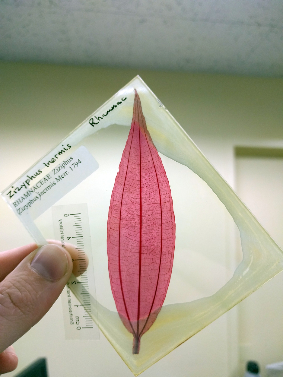
Herbaria are vital sources of comparative data for botanists and paleobotanists alike, especially those of us interested in patterns of leaf venation. Traditionally, one way paleobotanists have gotten images of leaf venation from herbarium sheets is to remove a leaf from the sheet, clear it until the tissue is translucent, and then stain it with safranin. Safranin is a dye that binds to the lignin in plant tissue, which makes the veins stand out against the leaf mesophyll. The stained leaves are then mounted on glass slides.
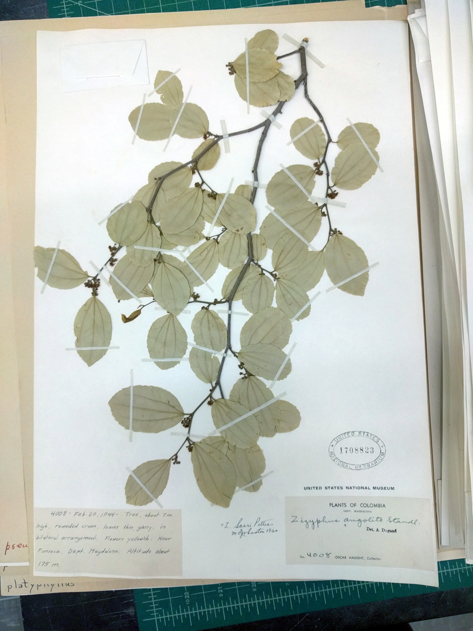
There are already large collections of cleared leaves around the world, including at the Smithsonian, Yale, Berkeley, and the Florida Museum of Natural History. Leaf clearing can give great results, but the process is slow and delicate: dried leaves are brittle to begin with, and they become flimsy and prone to tearing once they're cleared. Depending on the mounting medium, the finished slides may also need ongoing maintenance.
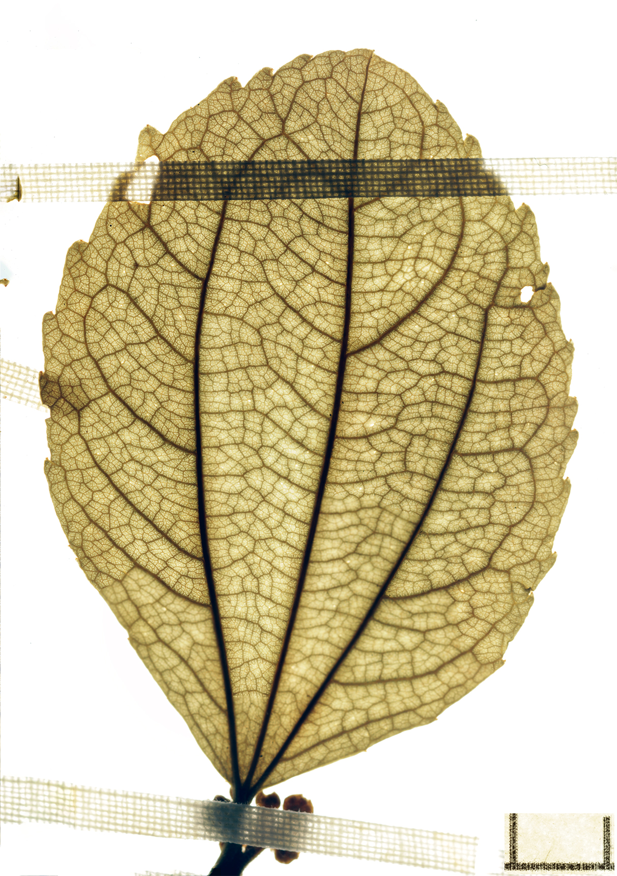
When I was working on Potomacapnos, I found that some of the leaves I wanted to compare weren't in the cleared leaf collections. I cleared some leaves myself, but I also experimented with other ways of imaging venation. To my surprise, the method that worked best was also the easiest: photographing leaves right on the herbarium sheet with a DSLR camera and a powerful flash from below. The light passes straight through most thin or moderately thin herbarium sheets, and through the leaf, sometimes with spectacular results (Jud and Hickey, 2013).
I recently visited the Smithsonian again to take new venation photos for an upcoming project. Here's one example of the results.
McNair (2015) has published a very similar approach, with more detail on the technique:
McNair, D. 2015. Imaging venation of broadleaf plants using DSLR cameras, transmitted light, and digital filters. Phytoneuron 39: 1–12.